Category: blog

  • Elevating Cardiovascular Care at Our State-of-the-Art Qoros Clear Lake Surgical Center

    Elevating Cardiovascular Care at Our State-of-the-Art Qoros Clear Lake Surgical Center

    Elevating Cardiovascular Care at Our State-of-the-Art Qoros Clear Lake Surgical Center

    In our pursuit of championing cardiovascular health, Bay Area Heart confronts the challenges posed by Peripheral Arterial Disease (PAD). This condition emerges from the insidious buildup of fatty deposits within arteries, constricting blood flow to the lower extremities. The consequences can be severe, ranging from discomfort and fatigue to heightened risks of heart attack, stroke, and even limb loss if left untreated.

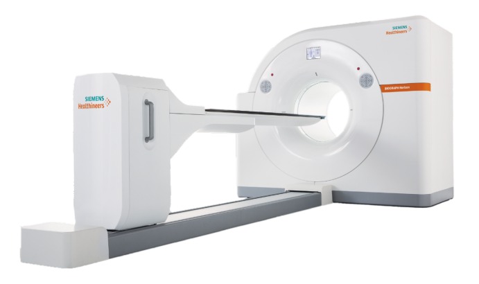
    Here at Bay Area Heart, we’re thrilled to unveil our latest endeavor in advancing cardiovascular care: the Qoros Clear Lake Surgical Center. Nestled within this state-of-the-art facility lies a hub of innovation, where we address not just PAD, but a myriad of cardiovascular conditions with precision and compassion.

    Early Detection: A Vital Step

    Our team of seasoned cardiologists stands ready to deploy cutting-edge technology and techniques to tackle a range of cardiovascular ailments, from PAD to coronary artery disease and beyond. Central to our approach is the early detection of conditions like PAD through comprehensive screening, particularly crucial for individuals harboring risk factors like smoking, diabetes, hypertension, and elevated cholesterol levels.

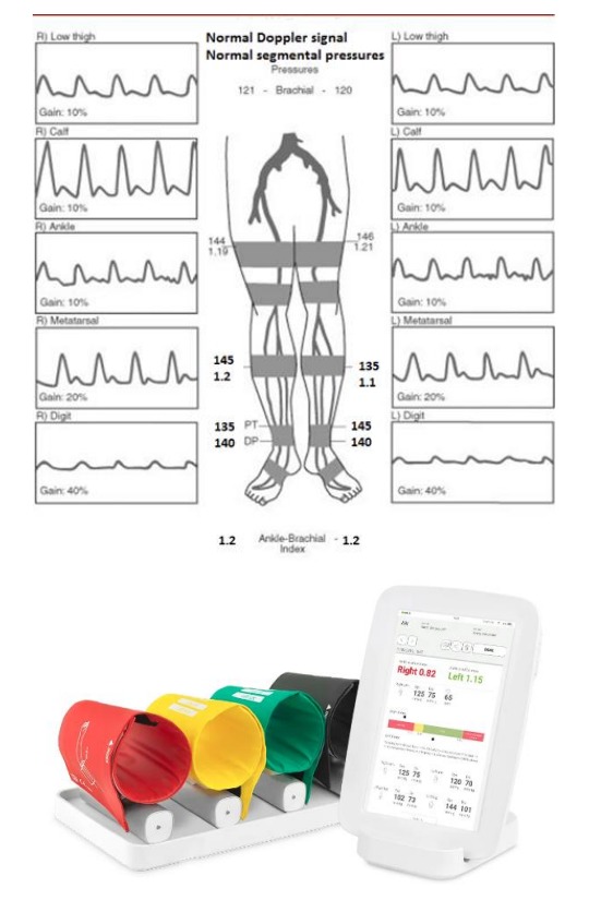
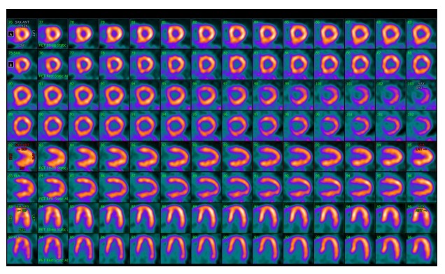
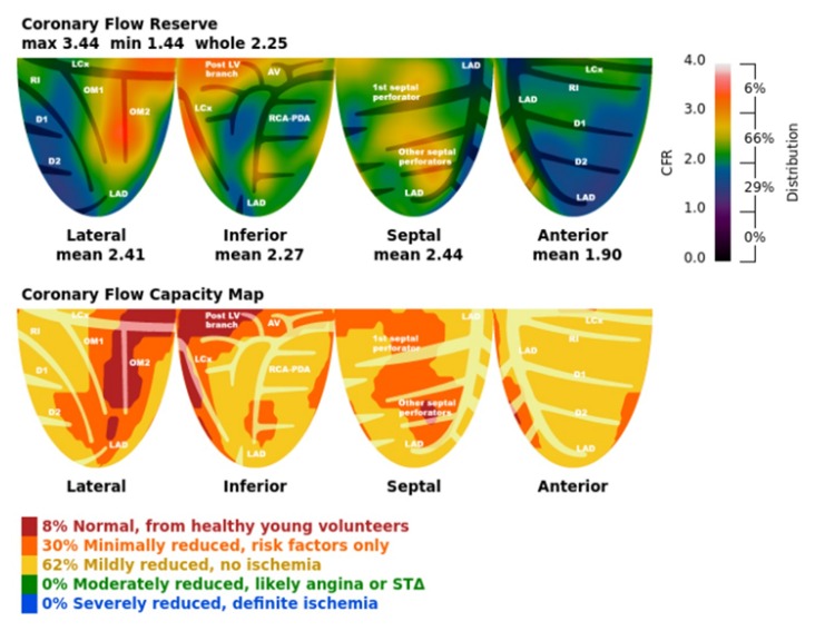
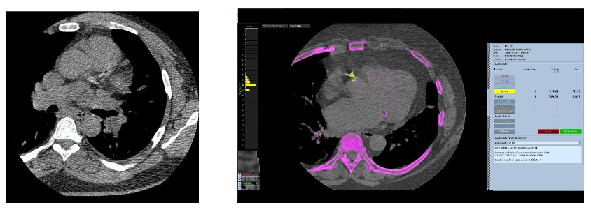
    Timely identification empowers us to initiate prompt interventions, potentially altering the course of diseases and preserving both lives and limbs. Screening protocols encompass a range of assessments, from physical examinations to specialized imaging studies such as ankle-brachial index (ABI) measurements and doppler ultrasounds.

    Innovative Treatment Modalities

    Once diagnosed, conditions like PAD open avenues for various treatment modalities, spanning lifestyle modifications, pharmacotherapy, and minimally invasive procedures like balloon angioplasty, stenting, and atherectomy. These interventions aim to enhance blood flow to the extremities, alleviating symptoms and enhancing overall health and mobility.

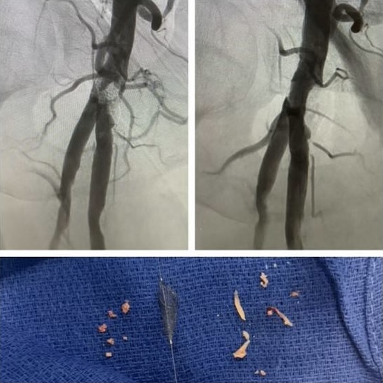
    Recently, our esteemed team achieved a significant milestone at the Qoros Clear Lake Surgical Center, successfully treating a patient afflicted with critical stenosis in the right common femoral artery. Employing a Fox Hollow’s SilverHawk atherectomy followed by drug-coated balloon angioplasty from Medtronic, our intervention yielded exceptional outcomes, marked by profound symptom alleviation and stellar final results.

    img

    A Beacon of Excellence in Cardiovascular Care

    This triumph exemplifies our proficiency in leveraging minimally invasive techniques within an outpatient setting, facilitating rapid recovery and same-day discharge for our valued patients. Under the expert guidance of Dr. Rakesh Shah, achieving over 25 years of experience in PAD management and interventional cardiology, the Qoros Clear Lake Surgical Center stands as a beacon of excellence in cardiovascular care.

    Equipped with state-of-the-art instrumentation and staffed by a compassionate, dedicated team, our facility epitomizes our unwavering commitment to delivering unparalleled care to our cherished community.

    Continuing the Journey Towards Health and Vitality

    For further insights into our services and capabilities, we invite you to explore our websites at Qoros Clear Lake ASC and Bay Area Heart. Together, let’s continue our journey towards safeguarding lives and limbs, one Texas heart at a time.

    In closing, we reaffirm the pivotal role of comprehensive cardiovascular care in preserving health and vitality, and at Bay Area Heart, we’re honored to be at the forefront of this life-saving mission.

    img
  • Peripheral Arterial Disease (PAD) and the  Importance of Screening

    Peripheral Arterial Disease (PAD) and the Importance of Screening

    Peripheral Arterial Disease (PAD) and the Importance of Screening

    What is Ankle Brachial Index (ABI)?

    Ankle Brachial Index (ABI) is a simple, non-invasive test that compares the blood pressure in your ankles to the blood pressure in your arms. This test is used to diagnose peripheral arterial disease (PAD), a condition in which the arteries in your legs or arms are narrowed or blocked. PAD can increase your risk of heart attack, stroke, and other serious health problems.

    How is ABI measured?

    The ABI test is performed by measuring the blood pressure in your arms and ankles using a blood pressure cuff and a Doppler ultrasound device. The ABI is calculated by dividing the systolic blood pressure in your ankle by the systolic blood pressure in your arm. An ABI of 1.0 to 1.4 is considered normal, while an ABI of less than 0.9 may indicate PAD.
    img

    How to interpret ABI results?

    1. An ABI of 1.0 to 1.4 is considered normal, while an ABI of less than 0.9 may indicate PAD. If your ABI is less than 0.9, your doctor may recommend further testing to confirm the diagnosis of PAD and determine the best course of treatment.

    2. ABI Interpretation:
         a. Normal ABI: 0.9 to 1.3
         b. Mild PAD: 0.7 to 0.9
         c. Moderate PAD: 0.4 to 0.7
         d. Severe PAD: Below 0.4

    What is Peripheral Vascular Disease (PVD)?

    Peripheral vascular disease (PVD) is a common condition that affects millions of people worldwide. It is caused by a buildup of fatty deposits in the arteries, which can reduce blood flow to the legs and feet. PVD can cause pain, cramping, and fatigue in the legs, and can increase the risk of heart attack and stroke.

    What devices are available for measuring ABI?

    There are several devices available for measuring ABI, including the MESI ABI system and the PADnet Express. These devices are designed to provide fast, accurate, and reliable measurements of ABI, helping doctors to diagnose PAD and monitor the effectiveness of treatment.

    How is PAD treated at Bay Area Heart?

    At Bay Area Heart, we are proactive in screening for and treating PAD. Our team of experienced cardiologists uses the latest technology and techniques to diagnose and manage this condition, helping our patients to reduce their risk of serious health problems and improve their quality of life
    img
  • Unlocking the Heart’s Secrets: Understanding Cardiac PET CT

    Unlocking the Heart’s Secrets: Understanding Cardiac PET CT

    Unlocking the Heart’s Secrets:
    Understanding Cardiac PET CT

    Dear Patients,

    When it comes to matters of the heart, technology continues to pave the way for more accurate diagnosis and personalized treatment. One such groundbreaking technology is Cardiac PET CT, a powerful imaging tool that offers unparalleled insights into the functioning of your heart. At Bay Area Heart, we believe in empowering our patients with knowledge about their health, which is why we are here to shed light on this remarkable diagnostic procedure.

    What is Cardiac PET CT?

    Cardiac PET CT, short for Positron Emission Tomography Computed Tomography, is a sophisticated imaging technique that combines the principles of PET and CT scans to provide detailed images of the heart. It allows healthcare providers to assess the blood flow to your heart muscle, detect any abnormalities, and evaluate the overall function of your heart.

    How Does it Work?

    During a Cardiac PET CT scan, a small amount of radioactive tracer is injected into your bloodstream. This tracer is specifically designed to highlight areas of your heart that may have reduced blood flow or metabolic activity. As you lie comfortably on the examination table, the PET CT machine captures images of your heart from different angles while the tracer circulates through your body.

    The CT component of the scan provides detailed anatomical images, allowing healthcare providers to precisely locate any areas of concern within your heart. By combining both functional and structural information, Cardiac PET CT offers a comprehensive view of your heart’s health.

    What to Expect During the Procedure?

    A Cardiac PET CT scan is a non-invasive procedure that typically takes around 30 to 60 minutes to complete. You will be asked to lie still on the examination table while the machine captures images of your heart. Throughout the process, our experienced technologists and healthcare providers will ensure your comfort and safety.

    Understanding the Results

    After your Cardiac PET CT scan, our team of experts will carefully analyze the images to assess the function and perfusion of your heart. The results provide valuable insights into the presence of any cardiovascular conditions, such as coronary artery disease or myocardial infarction. Based on these findings, we can develop a personalized treatment plan tailored to your specific needs.

    *Here is an example of a Cardiac PET CT image that showcases the intricate details of the heart’s anatomy and blood flow, allowing healthcare providers to pinpoint areas of concern with precision.*

    Example of a Cardiac PET CT Image

    img

    Example of a Coronary Flow Reserve Image

    img

    Image Source: Siemens Healthineers

    img3

    Bay Area Heart: Your Partner in Cardiovascular Health

    At Bay Area Heart, we’re committed to providing our patients with the highest standard of care using advanced technologies like Cardiac PET CT. Our team of board-certified cardiologists and specialists are dedicated to helping you achieve optimal heart health and well-being.

    Whether you are seeking preventive care, diagnostic imaging, or specialized treatment, you can trust Bay Area Heart to deliver compassionate and comprehensive cardiovascular services. With Cardiac PET CT, we’re able to offer cutting-edge diagnostic capabilities that empower you to take control of your heart health.

    If you have any questions about Cardiac PET CT or would like to schedule an appointment, please do not hesitate to contact us. Together, we can embark on a journey towards a healthier heart and a brighter future

    img
  • What is a Calcium Score and Why is it Important for Your Heart Health?

    What is a Calcium Score and Why is it Important for Your Heart Health?

    What is a Calcium Score and Why is it Important for Your Heart Health?

    What is a Calcium Score?

    A calcium score, also known as a coronary artery calcium (CAC) scan, is a non-invasive test that measures the amount of calcium in the walls of your coronary arteries. The coronary arteries are the blood vessels that supply oxygen-rich blood to your heart muscle. Calcium is a sign of plaque buildup, which can narrow or block your arteries and increase your risk of heart attack or stroke.

    What are Normal and Abnormal Values?

    A calcium score is expressed as a number that ranges from 0 to over 400. The higher the
    score, the more calcium and plaque you have in your coronary arteries. A score of:

    Calcium Score Risk Level
    0
    No identifiable plaque. Very low risk of heart disease.
    1-10
    Minimal plaque. Very low risk of heart disease.
    11-100
    Mild plaque. Low to moderate risk of heart disease.
    101-400
    Moderate plaque. Moderate to high risk of heart disease.
    Over 400
    Extensive plaque. High to very high risk of heart disease.

    What is the Testing Protocol?

    A calcium score test is done with a special type of X-ray machine called a computed tomography (CT) scanner. The test takes about 10 to 15 minutes and does not require any preparation. You will lie on a table that slides into the scanner, which will take pictures of your heart from different angles. You may be asked to hold your breath for a few seconds during the scan. The images will be analyzed by a computer and a radiologist, who will calculate your calcium score.

    What are the Patient Instructions?

    Before the test, you should inform your doctor if you are pregnant, have any metal implants, or have any allergies to contrast dye. You should also avoid caffeine and smoking for at least four hours before the test. You should wear comfortable clothing and remove any jewelry or metal objects from your chest area. After the test, you can resume your normal activities and diet. You should discuss your results with your doctor, who may recommend further tests or treatments based on your calcium score and other risk factors.

    How is the Interpretation Done?

    The interpretation of your calcium score is based on your age, gender, and ethnicity, as well as your personal and family history of heart disease, your blood pressure, cholesterol, blood sugar, and smoking status. Your doctor will use your calcium score and other information to estimate your 10-year risk of having a heart attack or stroke. Depending on your risk level, your doctor may advise you to make lifestyle changes, such as quitting smoking, eating a healthy diet, exercising regularly, and managing stress. Your doctor may also recommend that you repeat the calcium score test every few years to monitor your progress.

    Examples of Calcium Score Images

    Here are some examples of calcium score images from a CT scan. The white spots indicate the presence of calcium in the coronary arteries. The more white spots, the higher the calcium score and the higher the risk of heart disease.
    img

    Get Your Calcium Score Test at Bay Area Heart

    If you are interested in knowing your calcium score and your risk of heart disease, you can get your test done at Bay Area Heart, a leading cardiology practice in the Bay Area. We offer the calcium score test for only $99, which is much lower than the average cost of $200 to $400. We have a team of experienced and compassionate cardiologists who will guide you through the test and help you understand your results. We also provide personalized and comprehensive care for all your heart health needs. To schedule your calcium score test, call us at (832) 905-5940 or visit our website at www.bayareaheart.com.
    img